Membranes
I. Introduction
A. Why study membranes?
1. Membranes define and compartmentalize the cell
What part of the membrane is responsible for defining a compartment?
2. Membranes control the movement of substances
What part of the membrane is involved in transport?
3. Membranes are involved in the detection and recognition of signals
What molecule associated with a membrane is likely to be a receptor of a
signal?
4. The membrane is the locus of function in the cell
II. The Molecular Organization of Membranes
A. "average" composition:
52% protein
40% lipid
8% carbohydrate
B. Look at different membranes associated with specific cell
functions:
| Source | Protein % | Lipid % |
| Rat muscle | 65 | 35 |
| Rat liver | 60 | 40 |
| Rat liver mitochondrion | 70 | 27-29 |
| Human CNS myelin | 20 | 79 |
Note how the amount of protein parallels the diversity and degree of function
associated with that membrane.
C. The molecular components:
1. LIPIDS
-phosphatidyl choline (neutral lipids)
2. The Nature of Lipid Orientation
3. CARBOHYDRATES
4. PROTEINS
Integral (Intrinsic) 70% of total protein
Peripheral (Extrinsic) 30% of total protein
What is membrane protein classification based upon?
Peripheral
Integral
More detail on classification
Integral proteins are transmembrane proteins
integral proteins may be "singlepass" or. "multipass"
Peripheral proteins (3, 4 & 5) can be attached to the membrane by fatty
acids, the phospholipid, phosphatidyl inositol or by various non-covalent interactions
with other proteins.
example: "spectrin"
D. Models of Membrane Structure
1. Brief Historical Review
-oil-water partition coefficient
-Any theories to explain the results of Collander?
-Two postulates
a.lipid and integral proteins are disposed in a mosaic arrangement;
b.biological membranes are quasi-fluid structures in which both the lipids and integral proteins are able to perform movements within the bilayer.
2. Review questions:
-Explain how hydrophobic and hydrophilic regions of proteins and lipids orient themselves in a membrane.
-What determines the hydrophilic and hydrophobic regions of a protein? What environmental factors can affect these regions?
3. Evidence for a mosaic arrangement of proteins in the membrane
4. Evidence for membrane fluidity
5. Factors affecting membrane fluidity
membrane
fluidity can be thought of as equivalent to function
-unsaturated
-Any difference between cis and trans double bonds?
Membranes from plants and animals that live at low temperatures tend to have a higher concentration of unsaturated fatty acids than saturated fatty acids. Why?
-Which type of membrane (generally speaking) has a lower transition temperature: plant or animal? Any exceptions to this generalization?
E. Cell Membrane Permeability and Transport
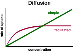
1. Types of movement through membranes:
Diffusion
Facilitated transport
Vesicle-mediated transport
2. Diffusion
Note that there is no standard free energy of formation, the substance does
not change from the initial to the final state; (delta)G0 = 0
where
J = flux density (molar cm sec-1)
D = diffusion constant (affected by molecular weight, mobility, temperature; D= U[mobility; size and shape]R[gas constant]T[temperature])
X = distance of diffusion (thickness of membrane).
Example problem:
calculate the flux density of sucrose moving across a 1 mm thick freely
permeable membrane of 1 cm2 area that separates a 1 M solution from a 0.1 M
solution.
So:
J = -D(delta)C/X
Dsucrose in water = 29 X 10-7 cm2/sec
(delta)C = 0.1 - 1.0 = -0.9 M (- = destination is a lower concentration)
X = 0.1 cm
J = - ( 29 X 10-7) -0.9/0.1 = 2.6 X 10 -5 molar cm sec-1
Problem to do:
Assuming the above concentrations and other conditions, calculate flux
density for urea (D = 109 X 10-7) and lysozyme (D = 10 X 10-7) and
explain the differences in the result.
Problems:
What if the thickness of the membrane is not known?
What if the substance is variably permeable through the membrane?
What about the consequence of "unstirred layers" near the surface
of the membrane?
permeability constant (P)
The "new" flux:
J = -P(delta)C
allows for comparison of different cell types, solutes and different
mechanisms of transport.
Example:
for tryptophan:
P = 10-7 cm/sec
assume the concentration gradient = -10-4 mol/sec
J = -10-7 cm/sec X (-10-4 mol/cm3) = 10-11
mol/sec through 1 cm2
2. Facilitated Transport
Two types:
carrier proteins
channel proteins
-carrier mediated transport will show saturation
The types of carrier systems:
uniport (single solute, one direction)
symport (two solutes, one direction)
antiport (two solutes, two directions)
-Glucose transport into blood cells from the extracellular fluid
-Do you think glucose will be permeable through the blood cell membrane?
-"ping" and "pong"
-movement is always downhill.
-show ion selectivity
-at high concentrations of ions can show saturation;
-pores are not continuously open; they have "gates" which open briefly and then close again;
-gates typically open in response to a specific perturbation of the membrane;
Examples: electrical excitability of nerve and muscle cells; leaf closing in Mimosa plants; cell motility in Paramecium
-polypeptide with 15 amino acids forms a coiled shape that inserts itself
into the membrane.
Can you predict how the amino acids of gramicidin will orient themselves in
the membrane? (They are: valine, glycine, alanine, leucine, alanine, valine, valine,
valine, tryptophan, leucine, tryptophan, leucine, tryptophan, leucine, tryptophan.)
Gramicidin is an antibiotic: how do you think it works?
Energetics of transport
uncharged solutes
charged solutes
uncharged: only concern is the concentration gradient across membrane; can
calculate "J" as discussed earlier or G.
charged solutes
need to take into account both concentration gradient and potential
(electrical charge) gradients found in cells.
What creates the electrical gradient?
Why is the electrical gradient important?
How do you calculate the level of the electrochemical gradient?
Example problem:
Calculate the free energy change that results from moving chloride (Cl-)
from the external solution to the cytoplasm of a cell at 37C. Assume that [Cl-]I
= 61 mM and [Cl-]o = 130 mM. The external electropotential
difference was measured to be 0.02 V. Remember that the ion does not change during
transport, so there is no standard free energy change (G0 = 0). Use the
Nernst-Planck Equation:
(delta)G = RT ln [Cl-]I/[Cl-]o +
zF(delta)Em
T = (37 + 273)K
R = 8.314 X 10⁻³ KJ/(mole K)
Z = -1 equiv/mole (equivalent of ion is the amount that has 1 mole of charge)
(delta)Em = membrane potential
F = Faraday constant (96.5KJ/(volt equiv mole)
(delta)G = (8.314 X 10⁻³)(310) ln 61/130 + (-1)(96.5)(-0.02)
= -0.02 KJ/mole
Primary active transport (transport protein that directly binds with ATP);
Secondary active transport (symport system where transport is driven by an ATPase generated ion gradient).
Example of primary active transport:
Na-K pump
Other points:
The Na-K pump is sensitive to certain plant derived steroids such as ouabain
What is the pump used for in the cell?
Ouabain has
been used as an "arrow poison". How do you think it works?
The sodium potassium pump has been experimentally demonstrated to be able to
work "backwards". What do you think would be the consequence of a backward
working pump? Would this pump produce any particular product?
Example of secondary active transport:
Epithelial cell
glucose absorption from intestine
3. Vesicle-mediated uptake
endocytosis
exocytosis
Focus on receptor-mediated endocytosis
1985: Goldstein and Brown (Nobel Prize)
-target cells have LDL receptors on membrane surface;
What kind of molecules are these receptors?
"coated pit" and clathrin
More about clathrin:
-"triskelion"